Amniotic Band Syndrome (ABS) is an uncommon congenital abnormality that causes entrapment of fetal parts (usually a limb or digits) in fibrous amniotic bands while in utero. It can result in disfigured feet. ABS affects about one in every 1,200 births and is believed to be the cause of 178 in 10,000 miscarriages. Some researchers believe that ABS is caused by early amniotic rupture, which leads to the formation of fibrous strands that entangle limbs and appendages. Treatment may include surgical excision of the fibrous band and any necrotic tissue.
Please contact our office to discuss your specific diagnosis and treatment options.
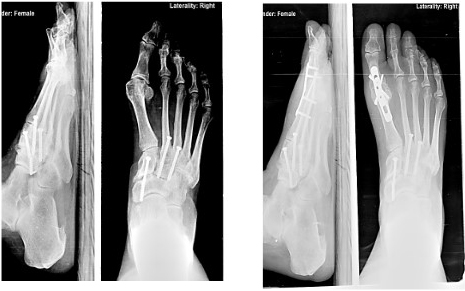
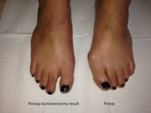
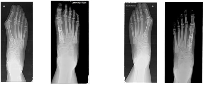
A bunion is a bone deformity caused by an enlargement of the joint at the base and side of the big toe (metatarsophalangeal joint). Bunions form when the toe moves out of place. The enlargement and its protuberance cause friction and pressure as they rub against footwear. Over time, the movement of the big toe angles in toward the other toes, sometimes overlapping a third toe (known as Hallux Valgus). The growing enlargement or protuberance then causes more irritation or inflammation. In some cases, the big toe moves toward the second toe and rotates or twists, which is known as Hallux Abducto Valgus. Bunions can also lead to other toe deformities, such as hammertoe.
Many people with bunions suffer from discomfort and pain from the constant irritation, rubbing, and friction of the enlargement against shoes. The skin over the toe becomes red and tender. Because this joint flexes with every step, the bigger the bunion gets, the more it hurts to walk. Over time, bursitis or arthritis may set in, the skin on the bottom of the foot may become thicker, and everyday walking may become difficult—all contributing to chronic pain.
Wearing shoes that are too tight is the leading cause of bunions. Bunions are not hereditary, but they do tend to run in families, usually because of a faulty foot structure. Foot injuries, neuromuscular problems, flat feet, and pronated feet can contribute to their formation. It is estimated that bunions occur in 33 percent of the population in Western countries.
Treatment for Bunions
Because they are bone deformities, bunions do not resolve by themselves. The goal for bunion treatment is twofold: first, to relieve the pressure and pain caused by irritations, and second to stop any progressive growth of the enlargement. Commonly used methods for reducing pressure and pain caused by bunions include:
- Protective padding, often made from felt material, to eliminate the friction against shoes and help alleviate inflammation and skin problems.
- Removal of corns and calluses on the foot.
- Changing to carefully fitted footwear designed to accommodate the bunion and not contribute toward its growth.
- Orthotic devices—both over-the-counter and custom made—to help stabilize the joint and place the foot in the correct position for walking and standing.
- Exercises to maintain joint mobility and prevent stiffness or arthritis.
- Splints for nighttime wear to help the toes and joint align properly. This is often recommended for adolescents with bunions, because their bone development may still be adaptable.
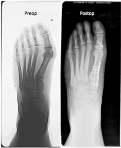
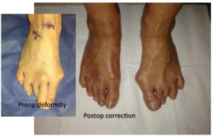
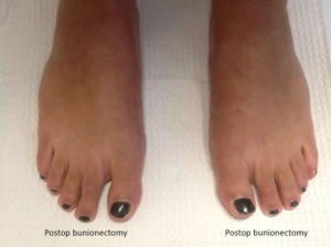
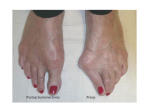
Surgical Treatment
Depending on the size of the enlargement, misalignment of the toe, and pain experienced, conservative treatments may not be adequate to prevent progressive damage from bunions. In these cases, bunion surgery, known as a bunionectomy, may be advised to remove the bunion and realign the toe.

Claw toe is caused by nerve damage from diseases like diabetes or alcoholism, which can weaken muscles in the foot. The term stems from the toes’ appearance—toes that look like claws digging down into the soles. Claw toe may lead to the formation of painful calluses. Claw toe worsens without treatment and may become a permanent deformity over time.
Common symptoms of claw toe include:
- Toes bent upward from the joints at the ball of the foot.
- Toes bent downward at the middle joints toward the sole of the foot.
- Corns on the top of the toe or under the ball of the foot.
Claw toe deformities are easier to repair when detected early. Splints or tape is used to hold the toes in correct position.
Clubfoot is one of the most common, non-life threatening, major birth defects among infants globally. Approximately one in every 1,000 newborns has clubfoot. Of those, one in three have both feet clubbed. The exact cause is unknown. Two out of three clubfoot babies are boys. Clubfoot is twice as likely to occur if one or both parents and/or a sibling has had it. Less severe infant foot problems are often incorrectly called clubfoot.
Clubfoot twists the heel and toes inward. It often appears like the top of the foot is on the bottom. Additionally, the clubfoot, calf, and leg are smaller and shorter than normal. When clubfoot is detected at birth, it is not painful and is correctable.
The goal of treating clubfoot is to make the infant’s clubfoot (or feet) functional, painless, and stable by the time he or she is ready to walk. Serial casting is the process used to slowly move the bones of a clubfoot into the proper alignment. The doctor starts by gently stretching the child’s clubfoot toward the correct position. A cast is put on to hold the foot in place. One week later, the cast is removed, the baby’s foot is stretched a little farther toward the correct position, and a new cast is applied. X-rays are used throughout the process to check on progress toward proper foot alignment. Casting generally repeats for 6-12 weeks and may take up to 4 months.
About half the time, clubfoot straightens with casting. Once the proper foot alignment is achieved, the child is fitted with special shoes or braces to keep the foot straight once corrected. These maintenance devices are used until the child has been walking for up to a year or more. Muscles for children with clubfoot commonly try to return to the clubfoot position; a regular occurrence among 2 and 3-year-olds, but a condition that may continue up to age 7.
In some cases, stretching, casting, and bracing is not enough to correct clubfoot. Surgery may be required to adjust the tendons, ligaments, and joints in the foot and ankle.
Corns are calluses that form on the toes because of bones that push up against shoes and build up pressure on the skin. The surface layer of the skin thickens, irritating the tissues underneath. Hard corns are usually located on the top of the toe or on the side of the small toe. Soft corns resemble open sores and develop between the toes as they rub against each other.
Improperly fitting shoes are a leading cause of corns. Toe deformities, such as hammertoe or claw toe, also can lead to corns. Self-care for corns includes soaking feet regularly and using a pumice stone or callus file to reduce the size of the corn. Special over-the-counter, non-medicated, donut-shaped foam pads can be worn to help relieve the pressure and discomfort. For large or lasting corns, please contact our office. We can shave off the corns using a scalpel.
Dysplasia, also known as epiphysealis hemimelica, is a disorder that affects the bone joints. It is characterized by overgrowth of the cartilage on the end of one or more of the long bones (carpal or tarsal bones) in the hand or foot. Usually, only one limb is involved. Dysplasia may cause limbs that are unequal in length. Less often, the cartilage on other bones, such as those in the ankle, knee, or hip joint, can be impacted.
Enchondromas are small benign tumors made up of cartilage that form in the bone beneath the toenail. Enchondromas are the most common bone tumors of the hands and feet and usually are painless. The tumor can involve large portions of the bones, causing thinning of the cortex. This can weaken the bone and cause it to break spontaneously. When enchondromas occur in the small bone in the end of the toe, they can cause pain that may mimic the pain of ingrown toenails.
Ollier’s Disease, also known as enchondromatosis, frequently occurs in the small bones in the hands and toes (phalanges) and the long bones behind the phalanges, called metatarsals. This condition is characterized by multiple enchondromas.
Maffucci’s Syndrome is a very rare form of enchondromatosis that combines multiple enchondromas in bones anywhere in the body with benign soft tissue tumors (known as hemangiomas), which are associated with blood vessels. This condition tends to appear in the hands and feet, and has a greater tendency toward malignant transformation than Ollier’s Disease.
The majority of enchondromas require no treatment. Only in cases where the tumors are aggressive and begin destroying bone tissue do they require further attention, often surgical removal.
Gordon Syndrome is an extremely rare disorder that belongs to a group of genetic disorders known as the distal arthrogryposis. These disorders typically involve stiffness and impaired mobility of certain joints of the lower arms and legs (distal extremities) including the knees, elbows, wrists, and/or ankles.
Joints affected by this disorder tend to be permanently fixed in a bent or flexed position. In the foot, Gordon Syndrome is characterized by the abnormal bending inward of the foot. The range and severity of symptoms may vary from case to case. Gordon Syndrome is believed to be an inherited condition.
Haglund’s Deformity (also known as pump bump or retrocalcaneal bursitis) is a painful enlargement on the back of the heel bone that becomes irritated by shoes. It normally appears as a red, painful, and swollen area in the back of the heel bone. Women tend to develop the condition more than men because of irritation from rigid heel counters in shoes (the piece of leather forming the back of a shoe) that rub up and down on the back of the heel bone.
Changing shoes, soaking feet, and anti-inflammatory medications often mitigate the symptoms of this problem. Note: Please consult your physician before taking any medications.
Hallux Limitus is a condition that results in stiffness of the big toe joint. It is normally caused by an abnormal alignment of the long bone behind the big toe joint, called the first metatarsal bone. Left untreated, Hallux Limitus can cause other joint problems, calluses, and/or diabetic foot ulcers. Painful bone spurs also can develop on the top of the big toe joint.
Anti-inflammatory medications, cortisone injections, and/or functional orthotics are some of the common treatments for stiff big toe. Note: Please consult your physician before taking any medications. Surgery may be required if spurring around the joint becomes severe.
Hallux Varus is a condition in which the big toe points away from the second toe. It is a possible complication from bunion surgery. The condition has been linked to a number of other causes, including congenital deformity, tight or short abductor hallucis tendons, trauma, injury, or an absence or surgical removal of a fibular sesamoid.
Treatment may focus on stretching the abductor hallucis tendon through specific exercises or toe splints. In severe cases, surgery may be recommended. During the surgery, a small incision is made on the side of the toe and the toe is splinted in a neutral or straight position.
Hammertoe is a deformity of the second, third, or fourth toes. In this condition, the toe is bent at the middle joint, causing it to resemble a hammer. Left untreated, hammertoes can become inflexible and require surgery. People with hammertoe may have corns or calluses on the top of the middle joint of the toe or on the tip of the toe. They may also feel pain in their toes or feet and have difficulty finding comfortable shoes.
Causes of hammertoe include improperly fitting shoes and muscle imbalance.
Treatment for the condition typically involves wearing shoes with soft, roomy toe boxes and toe exercises to stretch and strengthen the muscles. Commercially available straps, cushions, or non-medicated corn pads may also relieve symptoms.
In severe cases, hammertoe surgery may be recommended to correct the deformity.
Jackson-Weiss Syndrome (JWS) is a rare genetic disorder characterized by foot abnormalities. Symptoms include abnormally broad big toes, webbing of the skin between the second and third toes, an inward angling of the toes, and/or malformation or fusion of certain bones within the feet. Jackson-Weiss Syndrome is inherited and affects both sexes equally.
Mallet toes are deformities caused by bone and muscle imbalances that become exaggerated in people with active lifestyles. Arthritis can also lead to mallet toes. Mallet toes can cause extreme discomfort and may be aggravated if restrictive or improperly fitting footwear is worn for a prolonged period of time.
Treatment is designed to relieve pressure, reduce friction, and transfer forces from the sensitive areas. Shoes with a high and broad toe box (toe area) are recommended to prevent future irritation for people suffering from mallet toes. Other conservative treatments include forefoot supports, such as gel toe caps, gel toe shields, and toe crests. Gel forefoot supports provide immediate comfort and relief from common forefoot disorders without drying the skin.
Osteomyelitis is a type of bacterial bone infection that moves from acute to chronic phases quickly. The infection usually begins in another part of the body and spreads to the bone via blood. Traumatic injury, frequent medication injections, the use of a prosthetic device, and some surgical procedures can increase susceptibility to the underlying infection.
With osteomyelitis, the infected bone fills with a pus that deprives the bone of its needed blood supply. Over time, this can result in the death of bone tissue.
The presence of bone infection can be diagnosed with tests, such as bone scans and MRI. Osteomyelitis infections are very difficult to cure with oral or intravenous antibiotics. In chronic cases, surgical removal of the dead bone tissue is usually required.
The posterior tibial tendon starts in the calf, stretches down behind the inside of the ankle, and attaches to bones in the middle of the foot. This tendon helps hold the arch up and provides support when stepping off on your toes when walking. If it becomes inflamed, over-stretched or torn, it can cause pain from the inner ankle. Over time, it can lead to losses in the inner arch on the bottom of your foot and result in adult-acquired flatfoot.
Signs and symptoms of posterior tibial tendon dysfunction include:
- Gradually developing pain on the outer side of the ankle or foot.
- Loss of the arch and the development of a flatfoot.
- Pain and swelling on the inside of the ankle.
- Tenderness over the midfoot, especially when under stress during activity.
- Weakness and an inability to stand on the toes.
People who are diabetic, overweight, or hypertensive are particularly at risk. X-rays, ultrasound, or MRI may be used to diagnose this condition.
Left untreated, posterior tibial tendon dysfunction may lead to flatfoot and arthritis in the hindfoot. Pain can increase and spread to the outer side of the ankle.
Treatment includes rest, over-the-counter nonsteroidal anti-inflammatory drugs, and immobilization of the foot for six to eight weeks with a rigid below-knee cast or boot to prevent overuse. Note: Please consult your physician before taking any medications.
Bone spurs are a very common foot problem. In the feet, they develop most frequently in the heel, near the toes, and on top of the big toe joint. The spurs are small outgrowths of bone. In and of themselves, they are generally harmless. However, their location may cause friction or irritation from shoes or other foot structures, which can lead to other foot problems.
Heel spurs refer specifically to bone spurs in the heel. Heel spurs are growths of bone on the underside, forepart of the heel bone and occur when the plantar fibrous band pulls at its attachment to the heel bone. This area of the heel later calcifies to form a spur. With proper warm-up and the use of appropriate athletic shoes, strain to the ligament can be reduced.
Anti-inflammatory medications, cortisone injections, corrective shoes, and/or orthotics (special shoe inserts) are some of the common treatments for spurs. Note: Please consult your physician before taking any medication. Surgery may be prescribed if spurring around the joint becomes severe or leads to recurrent pain from persistent corns.
Tarsal coalition is a bone condition that causes decreased motion or absence of motion in one or more of the joints in the foot. The bones found at the top of the arch, the heel, and the ankle are referred to as the tarsal bones. A tarsal coalition is an abnormal connection between two of the tarsal bones in the back of the foot or the arch. This abnormal connection between two bones is most commonly an inherited trait.
The lack of motion or absence of motion experienced in a tarsal coalition is caused by abnormal bone, cartilage, or fibrous tissue growth across a joint. When excess bone has grown across a joint, it may result in restricted or a complete lack of motion in that joint. Cartilage or fibrous tissue growth can restrict motion of the affected joint to varying degrees, causing pain in the affected joint and/or in surrounding joints.
Symptoms usually include an aching sensation deep in the foot near the ankle or arch, accompanied by muscle spasms on the outside of the affected leg. Nonsurgical treatments, such as corrective shoes or custom orthotics, physical therapy, and anti-inflammatory medication, are the first courses of action. Note: Please consult your physician before taking any medications. Surgery is sometimes performed in severe cases to allow for more normal motion between the bones or to fuse the affected joint or surrounding joints.