Proudly Part of Privia Health
Glaucoma Drainage Implant Surgery
What are Glaucoma Drainage Implants?

Glaucoma drainage implants are small prosthetic devices that are placed to help lower the intraocular pressure and prevent further optic nerve damage. Glaucoma drainage implant surgery is an alternative to traditional filtration surgery (Trabeculectomy). In some patients, particularly those with certain types of glaucoma such as aphakic glaucoma, neovascular glaucoma, and uveitic glaucoma, trabeculectomies are known to be less successful at reducing intraocular pressure due to an aggressive healing response. Also, in patients who have had other eye surgeries, a glaucoma drainage device often works better than a trabeculectomy procedure to control the intraocular pressure. It should be noted that the glaucoma implant is not used to improve vision, but rather to lower intraocular pressure and prevent further vision loss from glaucoma. In this respect, this implant is completely different from the type of implant used during cataract surgery.
Glaucoma drainage implants are also successfully used as an initial surgical procedure for glaucoma. Various factors may influence the surgery recommended by your doctor. Sometimes an implant is necessary because there is expected to be extensive scarring in the outer layers of the eye. Compared to the channel made with trabeculectomy, the tube of a glaucoma implant is less likely to become blocked by this scar tissue.
How do drainage implants work?
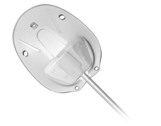
Glaucoma drainage implants come in different shapes and sizes. There are two general types of implants: Valved and Non-valved implants. All these implants have a tube and plate design. Regardless of which type of implant is used, a silicone tube is inserted into the front of the eye, usually between the cornea and iris, but other locations are occasionally used. The tube is like an artificial drain, allowing fluid to pass through it to a plate, which has been placed on the surface of the eye and acts as a reservoir.
The fluid then slowly percolates through this reservoir and is absorbed into the body fluids. The implant plate is usually placed in the area underneath the upper eyelid. Unless the lid is pulled back, neither you nor your family will notice it. With the upper lid retracted, a clear or white patch may be noted. This is a patch that covers the tube and prevents irritation. With all drainage implants, it can take 3 months or longer after surgery for the intraocular pressure to stabilize, as the capsule surrounding the plate of the implant needs time to mature in the eye.
What is my chance of success with after Glaucoma Drainage Implant Surgery?
Studies have shown that the success of glaucoma drainage implants is similar to those of trabeculectomy. It should be noted that glaucoma implants are sometimes used in patients with more complicated problems, and therefore the success rate in these patients may be lower than trabeculectomy in a standard eye. However, in many patients, these implants may be the best remaining available option. In about 5-10% of cases a second tube implant is necessary to adequately control intraocular pressure. When a second tube is necessary it is usually place in the lower part of the eye under the lower eyelid.
Remember that the goal of glaucoma implant surgery is to lower intraocular pressure and preserve vision. It will not restore vision that has already been lost. By lowering eye pressure, it is hoped that the operated eye will be spared further glaucomatous damage and can maintain its vision. As with any eye surgery, there is a risk of loss of vision, though this risk is low. Sometimes your doctor will combine the tube implant surgery with cataract surgery. In these cases there may be some visual improvement from clearing of the cataract and replacing it with a clear intraocular lens implant.
What is involved with a Glaucoma tube procedure?
After discussing the risk, benefits, and alternatives to surgery, your doctor will decide on the appropriate type of tube implant to be placed in your eye. When you and your doctor make a decision to proceed with placement of a glaucoma drainage implant, you will meet with our preoperative scheduler who will give you detailed instructions on how to prepare yourself for your upcoming surgery and what is involved in getting to the operating room for the procedure.
The surgery is an outpatient procedure performed in an ambulatory surgery center. In most cases, the surgery takes about one hour, though you will be at the surgery center for about 3-4 hours. The surgery is usually done under local anesthesia with intravenous sedation. An injection of local anesthetic numbs the eye completely so there is no discomfort and the eye will not move during surgery. Uncommonly a general anesthetic is used and the patient is put to sleep for the operation. Local anesthesia offers several advantages including less pain post-operatively, no sore throat from the airway tube used in general anesthesia, and quickly returning to normal alertness without the nausea often felt after general anesthesia. With local anesthesia, there is less risk than with a general anesthetic, especially in the elderly or those with health problems.
After surgery, the eye is covered by an eye patch and protected by a plastic shield overnight. On the morning following surgery, the patch/shield is removed and your ophthalmologist examines the eye. Eye drops are then used to prevent infection and reduce inflammation. It is important to take these as directed by your ophthalmologist since they can make a great deal of difference in the success of the procedure.
It may take several months after your surgery for the healing to be complete and for the implant to mature in your eye. During this time it is not unusual for your intraocular pressure, as well as your vision, to fluctuate. You will be ready to change your glasses prescription approximately 2-3 months after surgery.
